Glaucoma is the world's leading cause of irreversible blindness, affecting approximately 500,000 people in England and Wales and 80 million people worldwide.
Perimetry is the primary clinical method for identifying the condition but has largely remained unchanged since its inception over 40 years ago.
By drawing on 40 years of knowledge from basic science and clinical studies, we are investigating ways to detect glaucoma earlier, and identify its progression earlier (and with greater precision), than current methods. More accurate and precise measurements of changes in the visual field will not only enable more timely treatment to help prevent the progression of the condition, but it will also mean a reduction in the number of visual field tests that patients would be required to undergo, as well as reducing the burden on the health service.
The current glaucoma detection method
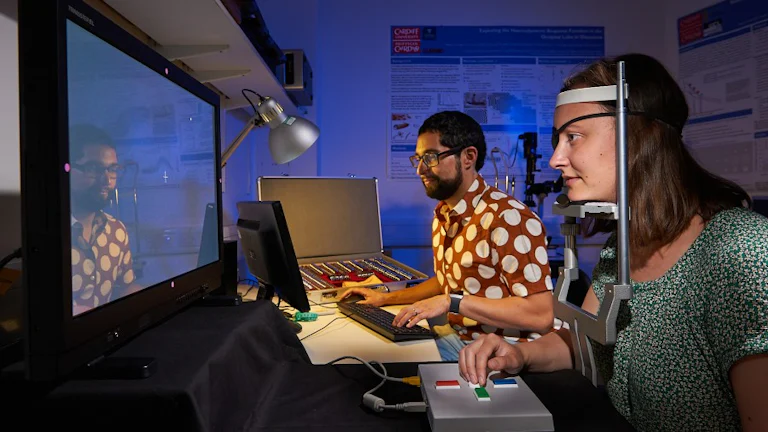
Perimetry is the name given to the measurement of the visual field. The current standard clinical test of the visual field involves asking patients to view a screen and press a button each time they see a small spot of light appearing briefly in their peripheral vision.
The objective is to find the dimmest spot that can be detected at multiple locations in the visual field and determine if visual field loss has occurred or progressed. However, the test design was based on legacy tests from over 40 years ago and is likely not optimal for accurate and precise measurements of changes in the visual field.
Our proposed method
Our novel method of testing the visual field will tap into biomarkers for glaucoma that our group more recently published.
Our research showed that the way the human eye and visual system gather light over a surface and over time is affected in early glaucoma. Tapping into these anomalies may enable earlier detection of subtle visual field changes for people with the condition or those at risk of it, as well as more precise determinations of progression over time.
This is especially important since earlier detection of glaucoma or its progression may lead to earlier and potentially more successful treatment.
Research
Our research draws on 40 years of basic science and clinical studies knowledge and brings collaborators from Wales, England and Northern Ireland. Our combined knowledge enables us to work towards a common goal of achieving more accurate and precise visual field testing for patients with and at risk of glaucoma.
Discoveries
In one of our research projects, we found that the way the human eye and visual system gather light over a surface (spatial summation) is affected in early glaucoma. Importantly, this was found in some areas of the visual field that could be considered ‘normal’ on a standard clinical visual field test.
In another study, we also found that how the eye and visual system gather light over time (temporal summation) is also affected in early glaucoma.
Our proposed alternative technique taps into these anomalies and promises to enable earlier detection and more precise measurements of subtle visual field changes.
View the papers:
- Sensitivity Loss in Early Glaucoma Can Be Mapped to an Enlargement of the Area of Complete Spatial Summation
- Spatiotemporal Summation of Perimetric Stimuli in Early Glaucoma
Research progress
Proof-of-concept
In 2018, we published proof-of-concept that our proposed glaucoma detection technique did outperform the current standard technique. The findings of that study, funded by the College of Optometrists, suggest that the new technique allows the detection of more subtle defects.
The results also showed that they may not be as variable as those in the current standard test in people with greater visual field loss. This could lead to more confident and timely decisions about whether or not an individual has glaucoma and whether or not established glaucoma is progressing.
Read more about the findings in our paper: Optimising the glaucoma signal/noise ratio by mapping changes in spatial summation with area-modulated perimetric stimuli
From labs to a clinical setting
In 2021, we received funding of over £1.82 million from the Medical Research Council (MRC) in the UK as part of its Developmental Pathway Funding Scheme (DPFS) to undertake further research to translate the test from university labs along the developmental pathway, to the clinical setting for patient benefit.
Next steps
This is our most ambitious research programme to date, and if successful, it could lead to significant improvements in the detection and monitoring of glaucoma in clinics worldwide.
We have strong links with the NHS in Wales, England, and the Health and Safety Executive (HSE) in Northern Ireland, and we have brought together an expert clinical and investigative team to conduct the study in three hospital sites. We also frequently engage with key stakeholders, including patients, clinicians, and industry representatives, to ensure that the needs of end users are prioritised in the research.
People
Principal Investigator
Co-Investigator
Post-doctoral Research Associate
Project Manager
Technology Transfer Officer
- Dr Matthew Stott
External Partners
- Dr Pádraig J Mulholland (Co-investigator, Ulster University)
- Professor Roger S Anderson (Co-investigator, Ulster University)
- Professor David F Garway-Heath (Co-Investigator, UCL)
- Dr Victoria Stapley (Post-doctoral Research Associate, Ulster University)
- Dr Wanghaoming Fang (Post-doctoral Research Associate, UCL)
Projects
Selected publications
REVAMP was born from a collaboration between the Principal Investigator and Co-Investigators, who have been working together in various vision science research projects since 2005. Here are some of our selected publications for projects our team have been involved with:
2024
- Anderson RS, Roark M, Gilbert R, Sumodhee D. Expert CONsensus on Visual Evaluation in Retinal disease manaGEment: the CONVERGE study. Br J Ophthalmol. 2024;31:bjo-2024-325310
- Montesano G, Crabb DP, Wright DM, Rabiolo A, Ometto G, Garway-Heath DF. Estimating the Distribution of True Rates of Visual Field Progression in Glaucoma. Transl Vis Sci Technol. 2024;13(4):15
- Thomas N, Acton JH, Erichsen JT, Redmond T, Dunn MJ. Reliability of gaze-contingent perimetry. Behav Res Methods. 2024;56(5):4883-4892
- Azuara-Blanco A, McCorry N, Tatham AJ, Georgoulas S, Founti P, Schweitzer C, Meier-Gibbons F, Denis P, Tuulonen A, Johannesson G, Martínez de la Casa JM, Prokosch V, Giannoulis DA, Abegão Pinto L, Garway-Heath DF, Topouzis F. European Glaucoma Society research priorities for glaucoma care. Br J Ophthalmol. 2024;108(8):1088-1093
2023
- Hunter AML, Anderson RS, Redmond T, Garway-Heath DF, Mulholland PJ. Investigating the Spatiotemporal Summation of Perimetric Stimuli in Dry Age-Related Macular Degeneration. Transl Vis Sci Technol. 2023;12(11):37
- Montesano G, Redmond T, Mulholland PJ, Garway-Heath DF, Ometto G, Romano D, Antonacci F, Tanga L, Carnevale C, Rossetti LM, Crabb DP, Oddone F. Spatial Summation in the Glaucomatous Macula: A Link With Retinal Ganglion Cell Damage.
Invest Ophthalmol Vis Sci. 2023;64(14):36 - Montesano G, Lazaridis G, Ometto G, Crabb DP, Garway-Heath DF. Improving the Accuracy and Speed of Visual Field Testing in Glaucoma With Structural Information and Deep Learning. Transl Vis Sci Technol 2023;12(10):10
- Montesano G, Mulholland PJ, Garway-Heath DF, Evans J, Ometto G, Crabb DP. Spatiotemporal summation of perimetric stimuli in healthy observers. J Vis. 2023;23(4):2
- Hunter AML, Anderson RS, Redmond T, Garway-Heath DF, Mulholland PJ. Investigating the linkage between mesopic spatial summation and variations in retinal ganglion cell density across the central visual field. Ophthalmic Physiol Opt 2023;(in press)
- Stapley V, Anderson RS, Redmond T, Saunders K, Mulholland PJ. Temporal summation in myopia and its implications for the investigation of glaucoma. Ophthalmic Physiol Opt 2023;43(4):788-797
2022
- Lazaridis G, Montesano G, Afgeh SS, Mohamed-Noriega J, Ourselin S, Lorenzi M, Garway-Heath DF. Predicting visual fields from optical coherence tomography via an ensemble of deep representation learners Am J Ophthalmol 2022;5:238:52-65
2021
- Patel DE, Cumberland PM, Walters BC, Abbott J, Brookes J, Edmunds B, Khaw PT, Lloyd IC, Papadopoulos M, Sung V, Cortina-Borja M, Raji JS, OPTIC Study Group. Study of optimal testing in children (OPTIC): developing consensus and setting reseach priorities for perimetry in the management of children with glaucoma. Eye (Lond) 2021:doi:10.1038/s41433-021-01584-0
- European Glaucoma Society Terminolgy and Guidelines for Glaucoma. 5th edition. Br J Ophthalmol 2021;105(Suppl 1):1-169
- Montesano G, McKendrick AM, Turpin A, Brusini P, Oddone F, Fogagnolo P, Perdicchi A, Johnson CA, Lanzetta P, Rossetti LM, Garway-Heath DF, Crabb DP. Do additional testing locations improve the detection of macular perimetric defects in glaucoma? Ophthalmology 2021;128(12):1722-1735
- Barkana Y, Leshno A, Stern O, Singer R, Russ H, Oddone F, Lanzetta P, Perdicchi A, Johnson CA, Garway-Heath DF, Rossetti LM, Skaat A. Visual field endpoints based on subgroups of points may be useful in glaucoma clinical trials: a study with the Humphrey Field Analyzer and Compass Perimeter. J Glaucoma 2021;20(8):661-665
- Gong Y, Zhu H, Miranda M, Crabb DP, Yang Haolan, Bi W, Garway-Heath DF. Trail-Traced Threshold Test (T4) With a Weighted Binomial Distribution for a Psychophysical Test IEEE J Biomed Health Inform 2021;25(7):2787-2800
2020
- Stapley V, Anderson RS, Saunders KJ, Mulholland PJ. Altered spatial summation optimizes visual function in axial myopia. Sci Rep 2020;10(1):12179
- Montesano G, Ometto G, Hogg RE, Rossetti LM, Garway-Heath DF, Crabb DP. Revisiting the Drasdo Model: Implications for Structure-Function Analysis of the Macular Region. Transl Vis Sci Technol 2020 14;9:15
- Founti P, Bunce C, Khawaja AP, Doré CJ, Mohamed-Noriega J, Garway-Heath DF, United Kingdom Glaucoma Treatment Study Group. Risk Factors for Visual Field Deterioration in the United Kingdom Glaucoma Treatment Study. 2020;127(12):1642-1651
- Wright DM, Konstantakopoulou E, Montesano G, Nathwani N, Garg A, Garway-Heath D, Crabb DP, Gazzard G; Laser in Glaucoma and Ocular Hypertension Trial (LiGHT) Study Group. Visual Field Outcomes from the Multicenter, Randomized Controlled Laser in Glaucoma and Ocular Hypertension Trial (LiGHT). Ophthalmology 2020;127(10):1313-21
- Montesano G, Rossetti LM, McKendrick AM, Turpin A, Fogagnolo P, Oddone F, Lanzetta P, Perdicchi A, Johnson CA, Brusini P, Garway-Heath DF, Crabb D. Effect of fundus tracking on structure-function relationship in glaucoma. Br J Ophthalmol 2020;104(12):1710-16
- King AJ, Hudson J, Fernie G, Burr J, Azuara-Blanco A, Sparrow JM, Barton K, Garway-Heath DF, Kernohan A, MacLennan G; TAGS Research Group. Baseline Characteristics of Participants in the Treatment of Advanced Glaucoma Study: A Multicenter Randomized Controlled Trial. 2020;213:186-94
2019
- Tribble JR, Vasalauskaite A, Redmond T, Young RD, Hassan S, Fautsch MP, Sengpiel F, Williams PA, Morgan JE. Midget retinal ganglion cell dendritic and mitochondrial degeneration is an early feature of human glaucoma. Brain Commun 2019;1(1):fcz035
- Gazzard G, Konstantakopoulou E, Garway-Heath D, Garg A, Vickerstaff V, Hunter R, Ambler G, Bunce C, Wormald R, Nathwani N, Barton K, Rubin G, Morris S, Buszewicz M. Selective laser trabeculoplasty versus drops for newly diagnosed ocular hypertension and glaucoma: the LiGHT RCT. Health Technol Assess 2019;23(31):1-102
- Gazzard G, Konstantakopoulou E, Garway-Heath D, Garg A, Vickerstaff V, Hunter R, Ambler G, Bunce C, Wormald R, Nathwani N, Barton K, Rubin G, Buszewicz M; LiGHT Trial Study Group. Selective laser trabeculoplasty versus eye drops for first-line treatment of ocular hypertension and glaucoma (LiGHT): a multicentre randomised controlled trial. Lancet 2019;393(10180):1505-16
2018
- Je S, Ennis FA, Woodhouse JM, Sengpiel F, Redmond T. Spatial summation across the visual field in strabismic and anisometropic amblyopia. Scientific Reports 2018;8(1):3858
- Rountree L, Mulholland PJ, Anderson RS, Garway-Heath DF, Morgan JE, Redmond T. Optimising the glaucoma signal/noise ratio by mapping changes in spatial summation with area modulated perimetric stimuli. Scientific Reports 2018;8(1):2172
- Yousefi S, Sakai H, Murata H, Fujino Y, Matsuura M, Garway-Heath D, Weinreb R, Asaoka R. Rates of Visual Field Loss in Primary Open-Angle Glaucoma and Primary Angle-Closure Glaucoma: Asymmetric Patterns. Invest Ophthalmol Vis Sci 2018;59(15):5717-25
- Garway-Heath DF, Zhu H, Cheng Q, Morgan K, Frost C, Crabb DP, Ho TA, Agiomyrgiannakis Y. Combining optical coherence tomography with visual field data to rapidly detect disease progression in glaucoma: a diagnostic accuracy study. Health Technol Assess 2018;22(4):1-106
- Garway-Heath DF, Quartilho A, Prah P, Crabb DP, Cheng Q, Zhu H. Evaluation of Visual Field and Imaging Outcomes for Glaucoma Clinical Trials (An American Ophthalomological Society Thesis). Trans Am Ophthalmol Soc 2017;115:T4
2017
- Matlach J, Mulholland PJ, Cilkova M, Chopra R, Shah N, Redmond T, Dakin SC, Garway-Heath DF, Anderson RS. Relationship between Psychophysical Measures of Retinal Ganglion Cell Density and In Vivo Measures of Cone Density in Glaucoma. Ophthalmology 2017;124(3):310-9
2015
- Mulholland PJ, Redmond T, Garway-Heath DF, Zlatkova MB, Anderson RS. Spatiotemporal Summation of Perimetric Stimuli in Early Glaucoma. Invest Ophthalmol Vis Sci 2015;56(11):6473-82
- Mulholland PJ, Redmond T, Garway-Heath DF, Zlatkova MB, Anderson RS. The Effect of Age on the Temporal Summation of Achromatic Perimetric Stimuli. Invest Ophthalmol Vis Sci 2015;56(11):6467-72
- Mulholland PJ, Zlatkova MB, Redmond T, Garway-Heath DF, Anderson RS. Effect of varying CRT refresh rate on the measurement of temporal summation. Ophthalmic Physiol Opt 2015;35(5):582-90
- Zhu H, Crabb DP, Ho T, Garway-Heath DF. More Accurate Modeling of Visual Field Progression in Glaucoma: ANSWERS. Invest Ophthalmol Vis Sci 2015;56(10):6077-83
- Garway-Heath DF, Crabb DP, Bunce C, Lascaratos G, Amalfitano F, Anand N, Azuara-Blanco A, Bourne RR, Broadway DC, Cunliffe IA, Diamond JP, Fraser SG, Ho TA, Martin KR, McNaught AI, Negi A, Patel K, Russell RA, Shah A, Spry PG, Suzuki K, White ET, Wormald RP, Xing W, Zeyen TG. Latanoprost for open-angle glaucoma (UKGTS): a randomised, multicentre, placebo-controlled trial. Lancet 2015;385(9975):1295-304
2014
- Mulholland PJ, Redmond T, Garway-Heath DF, Zlatkova MB, Anderson RS. Estimating the critical duration for temporal summation of standard achromatic perimetric stimuli. Invest Ophthalmol Vis Sci 2014;56(1):431-7
- Zhu H, Russell RA, Saunders LJ, Ceccon S, Garway-Heath DF, Crabb DP. Detecting changes in retinal function: Analysis with Non-Stationary Weibull Error Regression and Spatial enhancement (ANSWERS). PLoS One 2014;9(1):e85654
2013
- Redmond T, O'Leary N, Hutchison DM, Nicolela MT, Artes PH, Chauhan BC. Visual field progression with frequency-doubling matrix perimetry and standard automated perimetry in patients with glaucoma and in healthy controls. JAMA Ophthalmol 2013;131(12):1565-72
- Russell RA, Garway-Heath DF, Crabb DP. New insights into measurement variability in glaucomatous visual fields from computer modelling. PLoS One 2013;8(12):e8
- Redmond T, Russell RA, Anderson RS, Garway-Heath DF. Author response: Passing-Bablok regression is inappropriate for assessing association between structure and function. Invest Ophthalmol Vis Sci 2013;54(8):5850-1
- Redmond T, Anderson RS, Russell RA, Garway-Heath DF. Relating retinal nerve fiber layer thickness and functional estimates of ganglion cell sampling density in healthy eyes and in early glaucoma. Invest Ophthalmol Vis Sci 2013;54(3):2153-62
- Crabb DP, Smith ND, Glen FC, Burton R, Garway-Heath DF. How does glaucoma look?: patient perception of visual field loss. Ophthalmology 2013;120(6):1120-6
- Redmond T, Zlatkova MB, Vassilev A, Garway-Heath DF, Anderson RS. Changes in Ricco's area with background luminance in the S-cone pathway. Optom Vis Sci 2013;90(1):66-74
2012
- Russell RA, Crabb DP, Malik R, Garway-Heath DF. The relationship between variability and sensitivity in large-scale longitudinal visual field data. Invest Ophthalmol Vis Sci 2012;53(10):5985-90
- Russell RA, Malik R, Chauhan BC, Crabb DP, Garway-Heath DF. Improved estimates of visual field progression using bayesian linear regression to integrate structural information in patients with ocular hypertension. Invest Ophthalmol Vis Sci 2012;53(6):2760-9
2011
- Bergin C, Redmond T, Nathwani N, Verdon-Roe GM, Crabb DP, Anderson RS, Garway-Heath DF. The effect of induced intraocular straylight on perimetric tests. Invest Ophthalmol Vis Sci 2011;52(6):3676-82
2010
- Redmond T, Garway-Heath DF, Zlatkova MB, Anderson RS. Sensitivity loss in early glaucoma can be mapped to an enlargement of the area of complete spatial summation. Invest Ophthalmol Vis Sci 2010;51(12):6540-8
- Redmond T, Zlatkova MB, Garway-Heath DF, Anderson RS. The effect of age on the area of complete spatial summation for chromatic and achromatic stimuli. Invest Ophthalmol Vis Sci 2010;51(12):6533-9
2009
- Anderson RS, Redmond T, McDowell DR, Breslin KM, Zlatkova MB. The robustness of various forms of perimetry to different levels of induced intraocular stray light. Invest Ophthalmol Vis Sci 2009;50(8):4022-8
2006
- Anderson RS. The psychophysics of glaucoma: improving the structure/function relationship. Prog Retin Eye Res 2006;25(1):79-97
2002
- Garway-Heath DF, Holder GE, Fitzke FW, Hitchings RA. Garway-Heath DF, Holder GE, Fitzke FW, Hitchings RA. Invest Ophthalmol Vis Sci 2002;43(7):2213-20
- Ansari EA, Morgan JE, Snowden R. Glaucoma: squaring the psychophysics and neurobiology. Br J Ophthalmol 2002;86(7):823-6
2000
- Garway-Heath DF, Caprioli J, Fitzke FW, Hitchings RA. Scaling the hill of vision: the physiological relationship between light sensitivity and ganglion cell numbers. Invest Ophthalmol Vis Sci 2000;41(7):1774-82
Meet the team
Principal investigator

Academic staff

Dr Pádraig Mulholland
Honorary Research Fellow


Events
Speaking events
Our investigators regularly present novel research at national and international research conferences, including:
- Association for Research in Vision and Ophthalmology (ARVO)
- Imaging and Perimetry Society (IPS)
- North American Perimetry Society (NAPS)
- European Association for Vision and Eye Research (EVER)
- UK and Eire Glaucoma Society (UKEGS)
- World Glaucoma Congress (WCG)
- American Glaucoma Society (AGS)
Take part
How the study will work
People with glaucoma and people without glaucoma (control participants), of roughly the same age, will be invited to participate in a number of experimental tests, during which they will view a high-resolution screen and press a button every time they see a small spot of light appear on the screen in their side vision. We will compare the performance of new and current methods in terms of accuracy and precision.
Get involved
To help us develop the new test, we are inviting people with glaucoma, and people with no eye conditions, to come for some visual field tests. We are looking for participants who are aged over 40 years.
If you have glaucoma
We are recruiting people with either Primary Open Angle Glaucoma (POAG) or Normal Tension Glaucoma (NTG). If you would like to sign up to the study or would like further information, please contact us at VangasseR@cardiff.ac.uk.
If you have no eye conditions
We are also looking for a number of volunteers who do not have any eye conditions (though you may still be eligible if you wear spectacles or contact lenses for long/short-sightedness or for reading). If you are interested in participating or would like further information, please contact us at VangasseR@cardiff.ac.uk.
Locations
This study is being undertaken collaboratively by researchers at Cardiff University, Ulster University, and UCL. There will be three stages in the current project. In the current stage of the research, we would invite you to come for two visits within a 6-week period to one of the following sites (of your choice) for some short visual field tests:
- School of Optometry and Vision Sciences, Cardiff University, Cardiff
- NICRN (located within the Royal Victoria Hospital), Belfast Health and Social Care Trust, Belfast
- Moorfields Eye Hospital NHS Foundation Trust, London (from July 2023)
We will undertake some common clinical tests when you first arrive for your research appointment, to confirm that you are eligible to participate.
Please check back here for details of the next stages of the research, or feel free to contact us with an enquiry at VangasseR@cardiff.ac.uk.
Find out more about our research
We are recruiting people over 40 years old, with or without glaucoma to take part in our pioneering research.
Schools


Next steps
Research that matters
Our research makes a difference to people’s lives as we work across disciplines to tackle major challenges facing society, the economy and our environment.
Postgraduate research
Our research degrees give the opportunity to investigate a specific topic in depth among field-leading researchers.
Our research impact
Our research case studies highlight some of the areas where we deliver positive research impact.