We are working on all aspects of macular disease, but our primary focus is the development of new technologies that can detect and monitor sight threatening eye conditions at an early stage, before vision is lost.
Additionally, we are interested in the experiences of people with macular disease, with respect to their treatment, rehabilitation, and other aspects of their eye care.
About 196 million people in the world have age-related macular degeneration, the most common macular disease. It is the leading cause of visual impairment in the developed world. Typically, macular degeneration progresses slowly over the years from early-stage disease, when vision is largely normal, to advanced disease when vision is lost, sometimes abruptly. Currently, the only treatment available is for one form of advanced macular degeneration often referred to as ‘wet AMD’. This treatment, that involves injections into the eyeball, is not a ‘cure’ but it can delay further progression for some people.
We don’t think it makes any sense to wait for people to lose their eyesight before offering a treatment. By developing improved methods of detecting and monitoring early-stage disease and helping evaluate new therapies in clinical trials we are joining the global effort to stop people losing their sight to macular disease.
Aims
- To improve the diagnosis and monitoring of early-stage age-related macular degeneration.
- To evaluate new therapies for early-stage macular disease.
- To understand the relationship between early-stage macular disease and outer retinal function.
- To improve the lives and experiences of people living with advanced macular disease.
Research
Development of new diagnostics
We aim to improve the diagnosis and monitoring of early and intermediate stage macular disease by mapping the performance of a physiological process known as the ‘visual cycle’. The ‘visual cycle’ is responsible for the regeneration of visual pigment molecules after they have been deactivated following exposure to light and without it, we would all be blind in seconds.
Significantly, the visual cycle is dependent on the outer retinal complex, the same tissues that are affected in macular disease specifically, the choriocapillaris, Bruch’s membrane, the retinal pigment epithelium and photoreceptor outer segments. Hence, by measuring the visual cycle we are checking the health of the tissues that are directly affected by macular disease.
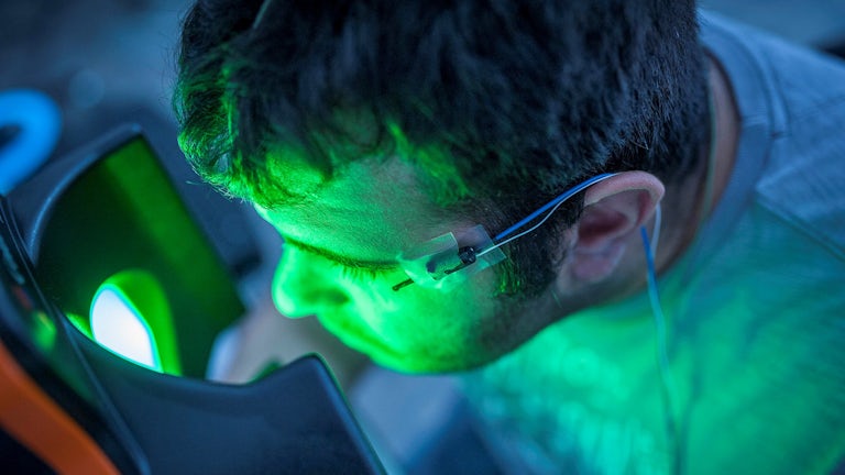
We measure the visual cycle using a new technique called high fidelity Imaging Retinal Densitometry (IRD). The technique has been developed in collaboration with the UK Astronomy Technology Centre and uses technology more usually associated with astronomy. The device and some typical results are shown in Figure 1.
Investigating experiences of patients with macular disease
In previous research, we have evaluated quality of life outcomes in individuals with macular disease and investigated the effectiveness of vision rehabilitation. Currently, we are researching patients’ experience of pain associated with eye injections to treat macular disease. In other research with the Ophthalmic Public Health group, we are using patient-reported experience measures to evaluate new eyecare pathways, including those for patients with age-related macular degeneration.
Projects
Saponins for Macular Disease (SAMADI)
This two centre, exploratory, randomised controlled trial is evaluating the feasibility of using oral glycoside supplements to treat people with early and intermediate stage age-related macular degeneration. The 30 month project is funded by AltRegen Ltd, coordinated by the Centre for Trials Research and led by Professor Tom Margrain.
Functional imaging of the outer retina using high fidelity imaging densitometry: exploring the relationship between visual pigment kinetics and AMD pathology
This project will use IRD to find out how drusen characteristics, including reticular pseudo drusen, effect visual pigment regeneration. We will test the hypothesis that cone pigment regeneration is selectively impaired by soft drusen and rod pigment regeneration by reticular pseudo drusen. This PhD project is funded by the College of Optometrists and is led by Dr Ashley Wood. The research student is Krishna Pattni.
Please read our participant information sheet to find out more.
High fidelity imaging densitometry: exploring the relationship between visual pigment kinetics, choroidal vasculature and sub-retinal drusenoid deposits in age-related macular degeneration
The aim of this study is to establish 1) the effect of age on IRD and OCTA results; 2) the relationship between outer-retinal function and choroidal vasculature in people with early stage AMD; 3) the relationship between IRD and conventional measures of dark adaptation and patient reported visual function. The PhD project is funded by the Macular Society and is led by Dr Ashley Wood. The research student is Vera Silva.
Please read our participant information sheet to find out more.
Investigation of pain associated with anti-vascular endothelial growth factor injections
In this project, we are evaluating patients’ experiences of pain during or after eye injections to treat age-related macular degeneration. We are investigating the factors that are associated with painful injections from both a patient and staff perspective. This PhD project is funded by the Abbeyfield Research Foundation and is supervised by Dr Ashley Wood and Dr Jennifer Acton. The research student is Christina Yiallouridou.

Find out more about our research
Our research delivers advances in knowledge to facilitate detection, diagnosis, monitoring and treatment of vision disorders to improve quality of life.
Images
Next steps
Research that matters
Our research makes a difference to people’s lives as we work across disciplines to tackle major challenges facing society, the economy and our environment.
Postgraduate research
Our research degrees give the opportunity to investigate a specific topic in depth among field-leading researchers.
Our research impact
Our research case studies highlight some of the areas where we deliver positive research impact.


