Cardiff leads way on lung ultrasound for management of Covid-19
13 May 2020
A Cardiff University-led review is being used globally to inform pioneering use of lung ultrasound to help in the management of patients with Covid-19.
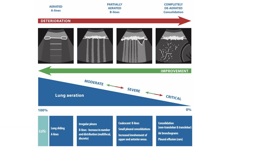
The academics were first to summarise early evidence on ultrasound imaging - more commonly used in pregnancy or for muscle injury - and how to use it to assess and monitor lung damage.
Lead author Dr Mike Smith, a senior lecturer from the University’s School of Healthcare Sciences, said: “As the storm clouds of the looming pandemic gathered, we very quickly collated the early evidence and guidance for use of lung ultrasound in Covid-19 patients.
“We wanted to take the information that was flying around the research and clinical communities and make it more useful for clinicians - right at the beginning of the pandemic curve. Since then lung ultrasound has emerged as critical to monitoring lung injury caused by Covid-19.”
Simon Hayward, a specialist physiotherapist from Blackpool Teaching Hospitals NHS Foundation Trust, said: “It’s an amazing tool because we can observe the changes in lung damage - and use it to inform how we manage these patients in real time. It has the potential to be integrated into care pathways for every Covid-19 patient with lung injury and to monitor their recovery.”
Lung damage is one of the most serious problems for patients with severe symptoms of Covid-19.
Key decisions such as admitting a patient to hospital, moving a patient from a ward to a high dependency unit, or whether they should be put on mechanical ventilation or taken off it, are usually informed by CT Scans, chest X-rays or by listening to the chest.
But for infection control reasons these cannot be used in the monitoring of Covid-19.
Co-author Dr Sue Innes, from the University of Essex, said: “Lung ultrasound plays an important part here as the standard assessment tool - the stethoscope - cannot be used with Covid-19 patients as it forms a direct connection between the patient's skin and the clinician's face. Lung ultrasound does not carry this risk and the infection control processes needed when using it are relatively straightforward.”
The researchers’ guidance was the first to collate how ultrasound imaging can be used to look at lung tissue in order to map deterioration or improvement.
The review also outlined how different healthcare workers could be trained in use of lung ultrasound to increase its availability. Usually, it is carried out by a relatively small but skilled workforce, comprised of intensivists (who specialise in care of critically-ill patients), anaesthetists, respiratory physiotherapists and advanced critical care practitioners and nurses.
The review identifies three workforces to upskill to bring about a 50-fold increase in availability.
They include clinicians who have previous experience in lung ultrasound; sonographers who already have high-level imaging skills but may have a lower than normal workload and experienced lung practitioners who don’t usually work with lung ultrasound.
“At a time of unprecedented pressure on healthcare services, supporting and educating clinicians is the key to unlocking the potential of lung ultrasound,” said Dr Smith.
The review was published in the journal Anaesthesia and co-authored by two expert NHS clinicians - Mr Hayward and Dr Ashley Miller from Shrewsbury and Telford Hospital NHS Trust.
Since its publication on 10 April it has received enormous worldwide attention, placing it among the top 0.2% of the 15 million scientific research papers ever tracked by Altmetric, which analyses the attention research outputs receive online.
Share this story
The School is dynamic, innovative and forward looking, and recognised for its excellence in learning, teaching and research.


